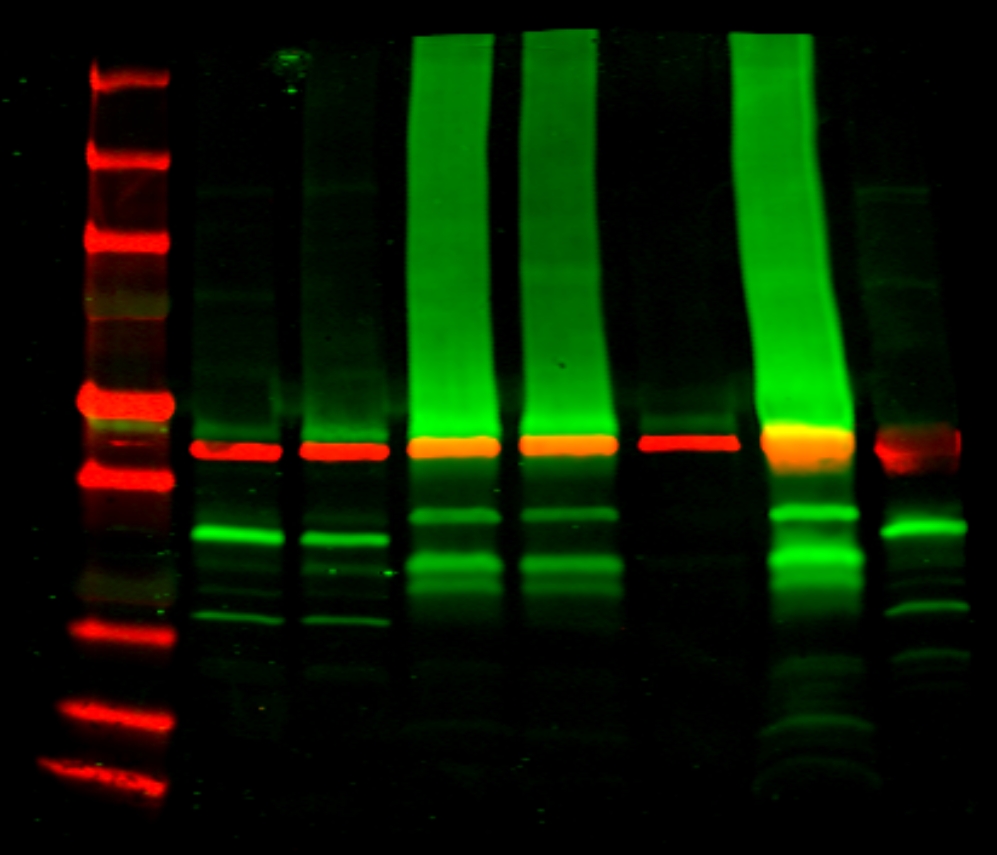
50 ug protein/well. We use 2 gels.
First Gel –> wells:
- Marker
- non-transfected HEK cells
- FLOT1/2-transfected HEK cells
- Rat brain
- Rat cerebellum
- Swine optic nerve
- Mouse brain
- SH-SY5Y (para comprovar que no tienen FLOT, porque las HEKs sin transfectar sí que tienen)
- non-transfected HEK cells
- FLOT1/2-transfected HEK cells
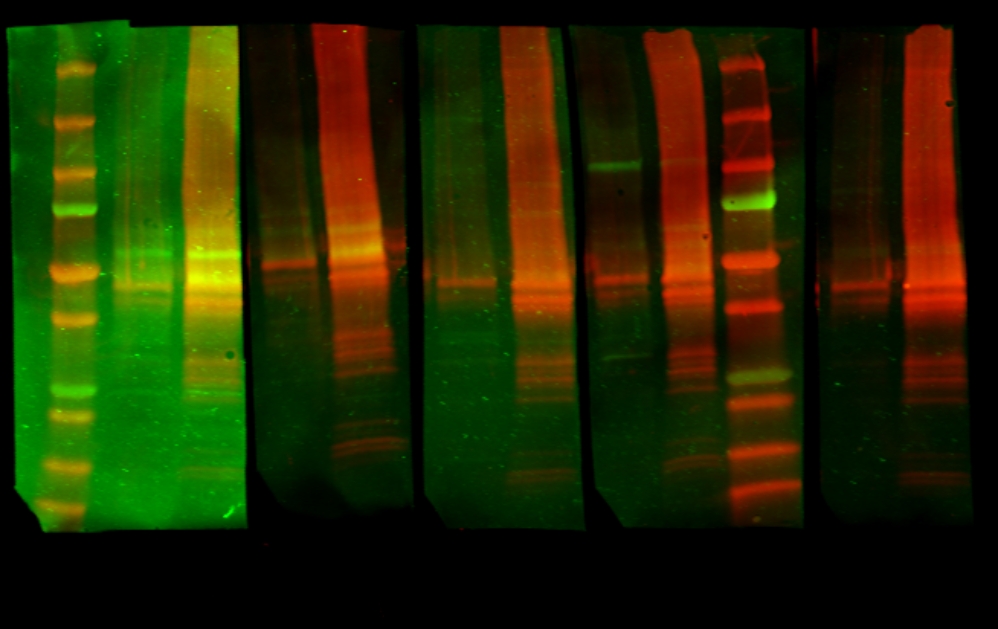
Second gel –> wells:
- Marker
- non-transfected HEK cells
- FLOT1/2-transfected HEK cells
- non-transfected HEK cells
- FLOT1/2-transfected HEK cells
- non-transfected HEK cells
- FLOT1/2-transfected HEK cells
- non-transfected HEK cells
- FLOT1/2-transfected HEK cells
- Marker
Protocol:
- We have used a precast gel (4-15% achrylamide) with 10 wells (Bio-Rad)
- Warm the samples at 96ºC for 5 minutes
- Load samples in the wells (30ul per well)
- Run with running buffer 1x (commercial –> BioRad) at 20 – 40mA
- Transfer to a nitrocellulose membrane with Transblot Turbo (using the commercial buffer) –> mixed proteins protocol
- Blotting: wash the membrane with water and cut it (in this case we have 6 membranes)
- Blocking: blocking buffer Casein – PBS 1:1 1h
- Primary antibodies (diluted in Casein-PBS 1:1) –> 2 hours at RT
- Mb1 (from well 1 to 8) –> Ac anti-FLOT1 (rabbit) 1/100 + Ac anti-B actin (mouse) 1/20.000
- Mb2 –> serum MS 158 1/50 + Ac anti-FLOT2 1/200
- Mb3 –> serum MS 163 1/50 + Ac anti-FLOT2 1/200
- Mb4 –> serum MS 247 1/50 + Ac anti-FLOT2 1/200
- Mb5 –> serum MS 316 1/50 + Ac anti-FLOT2 1/200
- Mb6 –> serum negative control 202-02 1/50 + Ac anti-FLOT2 1/200
- Wash with PBS-tween0’1% (3×5′)
- Secondary antibodies
- Mb1: GAR800 + GAM680 1/7500 (diluted in Casein-PBS tween0’1%): 1h at RT
- Mb2: GAH800 + GAR680 1/7500 (diluted in Casein-PBS tween0’1%): 1h at RT
- Wash with PBS-tween0’1% 1x (2×5′)
- Wash with PBS 1x (1×5′)
- Read with Odyssey equipment
RESULT:
- Mb1: la banda de FLOT1 (verde) se ve en todos los carriles menos en el de SH-SY5Y. Se observa más FLOT1 en los tejidos (a diferencia de lo que veíamos con FLOT2). Los lisados son correctos (la B-actina se ve bien). En las HEKs no transfectadas también hay FLOT1 y FLOT2.
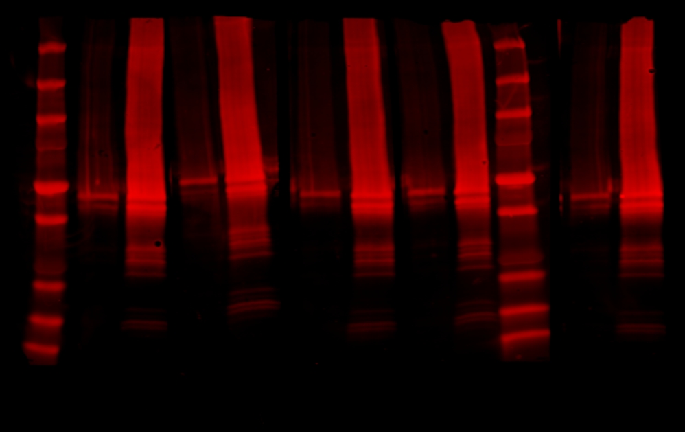
- Resto de membranas: hay mucho fondo rojo en los carriles con cels transfectadas FLOT1/2. Parece que se ve una banda en todos los pacientes que colocaliza con FLOT2, pero también parece que aparezca en el suero control negativo.