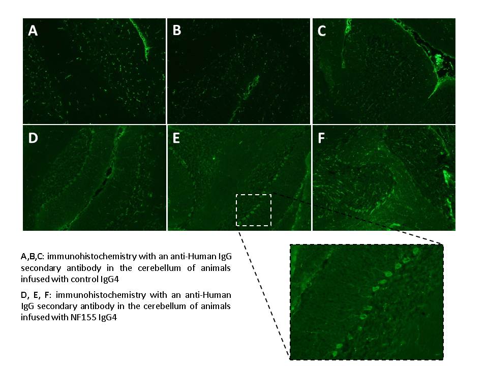
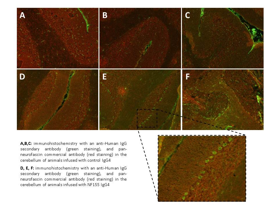
Samples:
- N31 (control)
- N33 (control)
- N34 (control)
- N35 (case)
- N36 (case)
- N37 (case)
Immunohistochemistry (in brain frozen sections):
- Fix with acetone (5 min)
- Wash with PBS 1x (3 x 5 min)
- Block with Blocking Goat (10 ml PBS + 0’5 ml Goat Serum + 0’2 g BSA) (1 h)
- Primary antibody: anti-NF. Diluted 1/300 in blocking goat (1h)
- Wash with PBS 1x (3 x 5 min)
- Secondary antibodies (1 h): GAH488 + GAC594 diluted 1/1000 in blocking goat (1h)
- Wash with PBS 1x (3 x 5 min)
- Fluoromount mounting medium
RESULTS:
- The pan-neurofascin doesn’t stain any specific structure.
- In the 3 cases there is a green staining in Purkinje cells (in comparison with the 3 controls) –> the human Ig is detected.