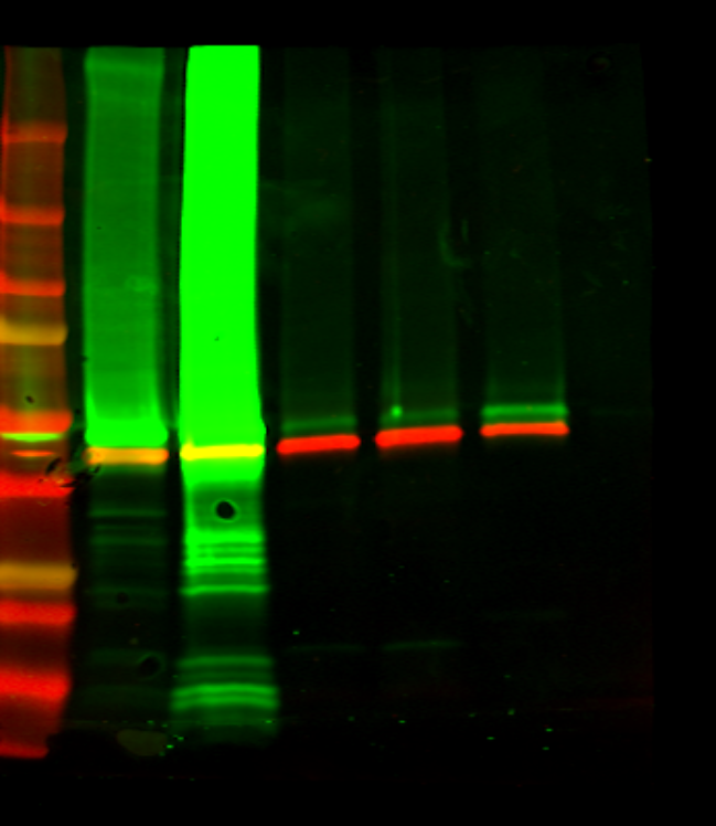
Wells:
50 ug protein/well
- Marker
- non-transfected HEK cells
- FLOT1/2-transfected HEK cells
- Rat brain
- Rat cerebellum
- Swine optic nerve
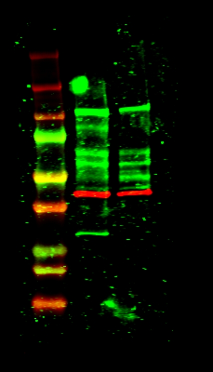
- –
- Marker
- non-transfected HEK cells
- FLOT1/2-transfected HEK cells
Protocol:
- We have used a precast gel (4-15% achrylamide) with 10 wells (Bio-Rad)
- Warm the samples at 96ºC for 5 minutes
- Load samples in the wells (30ul per well)
- Run with running buffer 1x (commercial –> BioRad) at 20 – 40mA
- Transfer to a nitrocellulose membrane with Transblot Turbo (using the commercial buffer) –> mixed proteins protocol
- Blotting: wash the membrane with water.
- Blocking: blocking buffer Casein – PBS 1:1 1h
- Primary antibodies (diluted in Casein-PBS 1:1) –> overnight at 4ºC:
- Mb1 (from well 1 to 6) –> Ac anti-FLOT2 (rabbit) 1/200 + Ac anti-B actin (mouse) 1/20.000
- Mb2 (from well 8 to 10) –> serum MS 316 (positive for FLOT1/2 by ICC) 1/50 + Ac anti-B actin (mouse) 1/20.000
- Wash with PBS-tween0’1% (3×5′)
- Secondary antibodies
- Mb1: GAR800 + GAM680 1/7500 (diluted in Casein-PBS tween0’1%): 1h at RT
- Mb2: GAH800 + GAM680 1/7500 (diluted in Casein-PBS tween0’1%): 1h at RT
- Wash with PBS-tween0’1% 1x (2×5′)
- Wash with PBS 1x (1×5′)
- Read with Odyssey equipment
RESULT:
- Mb1: La B-actina se ve bien en todos los pocillos (lisados bien hechos). Se ve en todos una banda verde a aprox 50 kDa, que podría ser la FLOT2 –> hacer el mismo WB pero con FLOT1 para comprovar
- Mb2: en el pocillo de HEKs transfectadas se ve una banda a aprox 50 kDa que no se observa en el pocillo de HEKs no transfectadas. Podría ser la FLOT del paciente.